Patient Case Study: 3D Printed Nose Bolus
University of Chicago
The following case demonstrates the application of technology used in clinical radiation oncology through the creation of simple bolus for electron radiotherapy. This case is a great example of how Adaptiiv’s software can be used to create a custom-shaped bolus around the nose.
Description
This case study demonstrates a clinical application of 3D printing in clinical radiation oncology by designing and manufacturing a bolus to treat a 57-year-old female with basal cell carcinoma (BCC) of the right nose. The workflow plan was to do a mini CT scan used to generate the bolus and then do a full CT simulation with the 3D printed bolus on the patient’s right nose and treat her disease with one electron field. The bolus application was necessary to deliver the prescribed dose to the skin surface.
Patient History
A 57-year-old female was treated at the University of Chicago Comprehensive Cancer Center at Silver Cross Hospital to manage basal cell carcinoma after Mohs micrographic surgery to the right nose. Her treatment options included a skin graft procedure or radiation therapy; the patient elected for the latter.
Fabrication and Treatment
The bolus was designed to achieve optimal dose distribution to the small target volume in the right nose and spare tissues distal to it. The width of the bolus was intended to cover the GTV with 20mm margins. A uniform bolus thickness of 7mm was used. DICOM images of the bolus were converted to a stereolithography file. The end product was fabricated with the Airwolf 3D AXIOM 20 M printer, using Cheetah™ by NinjaTek™ TPU filament. The printing duration was approximately 3 hours. The patient received 45Gy over 15 fractions at 6 MeV.
Summary
- The Adaptiiv software solution was used to create a patient-specific bolus with superior fit compared to a wax bolus.
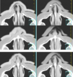
- 3D printing was used to create a custom-shaped bolus to follow surface irregularities around the nose and fill in the air space, reducing air gaps to no larger than 2mm.
- The bolus could be reproducibly placed at the times of planning CT and daily treatments, improving patient experience and comfort.
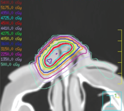
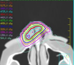
- An acceptable treatment plan was obtained (90% isodose to 100% of GTV).

Left side, axial CT slices superior-inferior of the right nose with the 3D printed bolus. Right side, corresponding CT slices superior-inferior of the right nose with the wax bolus.Left side, axial CT slices superior-inferior of the right nose with the 3D printed bolus. Right side, corresponding CT slices superior-inferior of the right nose with the wax bolus.

Acceptable treatment plan with the 3D printed bolus to the right nose.